
Central Venous Access
Warning
Objectives
To outline the expected conduct for placement of central venous access lines in the operational environment.
Scope
What are the indications for central venous line placement
What are the differences between elective versus emergency placement
What equipment is needed
What technique to use - US guided vs landmark
How to confirm placement
What documentation is required
What training and competence is required
What ongoing care of the line is required
What to consider if complications occur
Note - these guidelines are to support established clinical practice in the deployed setting. It is not an appropriate skill to learn on deployment.
Audience
This guideline is intended for use by registered healthcare professionals fulfilling a specialist role in forward medical locations, emergency department or deployed hospital settings.
Anaesthetists
Peri-operative Practitioners
Emergency Medicine Physicians
Emergency Department Nurses
Critical Care Nurses
Initial Assessment & Management
Indications for placement of Central Venous Line
Central Venous Access is gold standard for rapid infusions of large volume in hypovolaemia
- Most patients arriving at Role 2 will have IV or IO access
- Central venous access is to be considered at Role 2 and above.
- Central venous access requires suitably trained clinicians and may take time
- Large bore IV or IO access is considered first line
- Consider central venous access if rapid transfusion is required or peripheral IV access attempts have failed.
Prepare equipment and team before casualty arrives
- If there are elements of ATMIST that predict need for central venous access?
- Will CVP be required in an emergency?
- Is there time for elective placement utilising Ultrasound (US) or is landmark technique required?
- Can a second clinician place the CVP line concurrent with ongoing resuscitation?
Emergency Placement of CVP line
Placement during ongoing resuscitation
- Communication with team is essential to assist timing of insertion to prevent delays to other aspects of resuscitation of casualty.
- Attempt sterility as able.
Subclavian venous access using a landmark technique is ideal
- Access to the subclavian vein allows rapid placement of central venous access while airway management proceeds in tandem
- The subclavian vein is tented open by the clavicle and is easier to access than jugular veins in severe hypovolaemia
The subclavian ARTERY may be cannulated in error
- Consider this possibility until correct placement is confirmed
- Confirmation by arterial gas analysis may be difficult in extremis
Infusion through the subclavian artery may be life saving but infusion of medication before confirmation of venous placement must be avoided.
Clinicians should take the opportunity to learn/maintain landmark access skills prior to deployment. This is currently covered on the MOSTT course.
Elective placement of CVP
Ultrasound is available on operations in Role 2 and 3
- NICE guidance suggests that US is considered for all electively placed lines
- When time permits consider using safety checklists such as that provided by FFICM (in accordion section below)
Confirmation of placement
Line placement should be confirmed prior to use. This may be performed by:
1. Clinical acumen - presence and appearance of blood in syringe (colour maybe unreliable in severe shock)
2. Use of ultrasound (where time allows) - confirm intravascular wire placement prior to dilatation of vessel.
3. Arterial blood gas analysis - recommended if arterial cannulation is possible/likely.
4. Radiography - useful to check line tip position and to exclude other complications.
Advanced Assessment & Management
Complications of CVP access
Management of lines placed in Arteries
- When using US it is important to visualise the wire in the vessel prior to dilatation.
- Lines inserted without US or in cases of uncertainty blood gas analysis and radiology will aid confirmation of placement.
- Confirmation of venous placement MUST proceed prior to infusion of medication.
- Lines placed in arteries may be used with care for fluid resuscitation
- Documentation of complications and hand-over to other HCPs MUST occur
- Discussion with vascular surgeons is required in the case of arterially placed lines and this may require surgical or interventional radiology for removal.
Extravascular placement
Pneumothoraces
There is a significant risk of pneumothorax in particular with subclavian vessel access with the landmark technique.
- Clinicians should remain vigilant to this complication
- Raised intra-thoracic pressure
- Sudden cardiovascular collapse on commencement of mechanical ventilation
- Be prepared to decompress chest/insert intercostal drains.
- In addition line placement may damage nearby structures, or enter other spaces
-
- Pericardium - often fatal and may occur late due to line migration
- Pleural or peritoneal spaces - resulting in effusions from infused fluids.
Infection
- Ideally all lines are placed using aseptic precautions
- When this has not been possible consideration of line removal/change should take place once the patient is stable and in the critical care setting
- Line access points should be assessed regularly for signs of infection (VIP scores)
Failure to achieve Central Venous Access
- In cases of extreme hypovolaemia, central access may be challenging.
- Resuscitation should proceed in parallel with efforts to achieve central access if adequate personnel allow. This may necessitate access via other means for example intra-osseous. Beware becoming fixated and distracted on central access - the team leader must be vigilant to this.
- It may be prudent in certain situations to abandon efforts at central cannulation to allow for reversal of hypovolaemia via other means and to prevent damage to vasculature that may prevent further attempts.
- In the setting of prolonged casualty care where vasopressors may be required peripheral agents including norepinephrine may be used with care. See vasopressor and inotropes CGO
Documentation
Line insertion is to be documented on FMED.
Monitoring
Good practice suggests that all central lines are pressure monitored continually. Where there is suspicion of misplacement, arterial gas analysis may be useful.
Insertion sites should be regularly monitored using VIP (Visual Infusion Phlebitis) scores for signs of infection.
Score 0 - insertion site appears healthy - no signs of phlebitis. Continued observation of line site is indicated.
Score 1 - one of the following - slight pain or redness. Continued observation of line site is indicated.
Score 2 - two of the following - pain at site, redness or swelling. This is early phlebitis - consider changing line.
Score 3 - all the following- pain, redness and swelling. Medium stage of phlenitis - line should be changed.
Score 4 - all the following present and evident - pain, swelling, redness and palpable venous cord. Advanced phlebitis/start of thrombo-phlebitis. Replace line and consider starting treatment.
Score 5 - all the following present, evident and extensive - pain, redness, swelling, palpable venous cord, pyrexia. Advanced thrombo-phlebitis. Replace line and start treatment.
Regular audit
It is good practice to audit placement and management of central venous lines in the deployed setting. This should include indications, placement, complications, removal and link in with Role 4.
Prolonged Casualty Care
Should rapid evacuation to a higher level of care not be possible, consider;
Replacement of lines inserted in dirty environment
- All lines that have been put in where sterility has not been possible (during emergency resuscitation) or where sterility may not be confirmed (line placed in another facility) should be considered for replacement.
- Replacement of the line should be undertaken when the patient is stable and in a sterile manner.
- Consideration of pros and cons may favour leaving a line in place. Where this happens full documentation of decision making should take place.
- VIP scores and assessment of line infections should be regularly undertaken.
- Re-wiring a line should only occur if alternative access cannot be achieved.
- Use for medications in critical care should follow established critical care guidelines and practices.
Consideration and practice of removal of line
- At least daily the ongoing requirement for central access should be assessed and decision making documented.
- Line removal should follow established practices - it may be difficult in the deployed setting to achieve a head down posture to prevent air emboli - therefore this particular complication should be considered.
- Complications of line removal
- Vascular damage
- Air embolism
Paediatric Considerations
Central venous access on deployment is possible - 8cm,5.5 French gauge lines are in the module.
Transducers for the lines are the same as in adult practice so need to add 3ml/hr to fluid balance.
"Hockey stick" probes are not available on the Sonosite US - use the linear probe that is available.
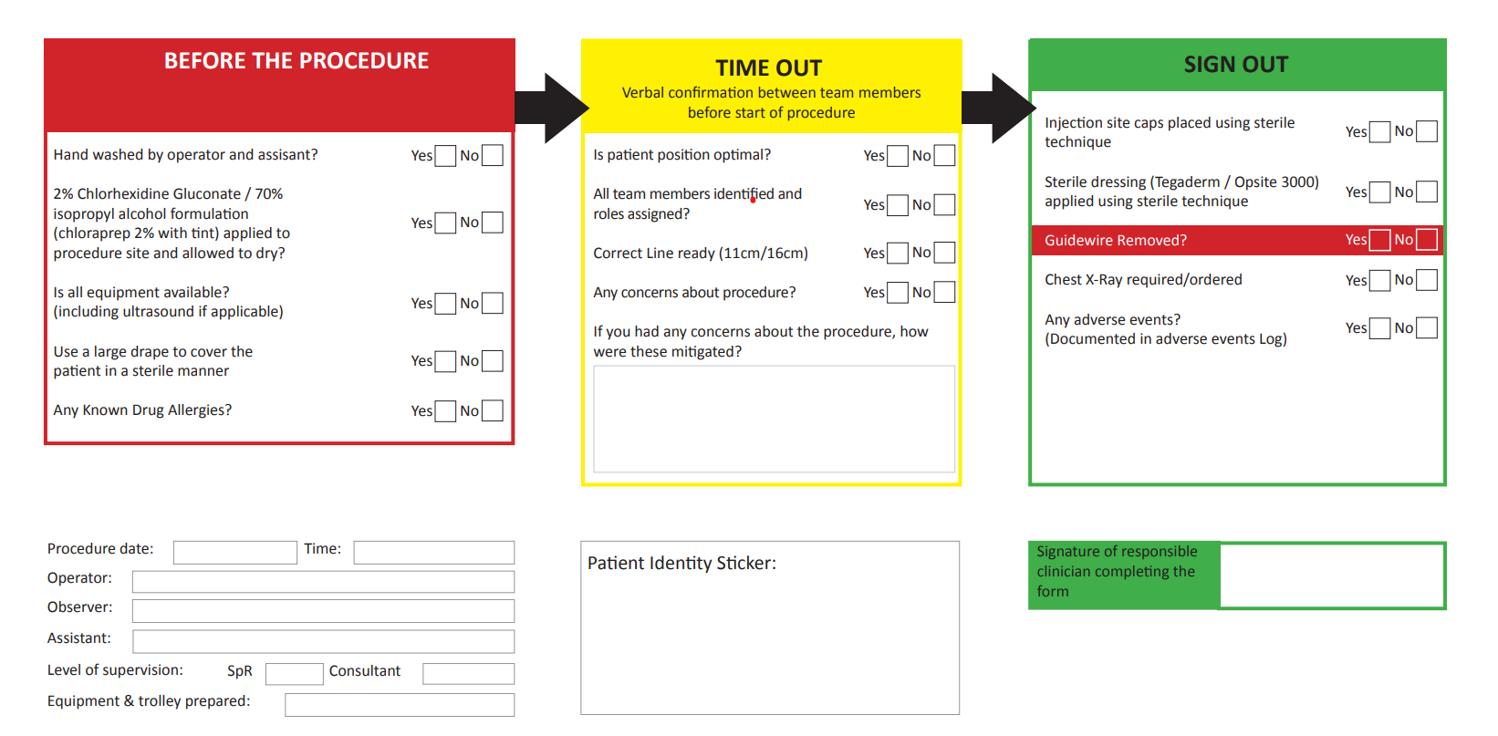
Central Venous Access Documentation - consider printing and adding to notes:
During the procedure (circle appropriate option)
Sterile gloves and sterile gown worn by operator and assistant Yes No
Hat and mask worn by operator and assistant Yes No
Sterile field maintained Yes No
Sterile sheath and sterile gel used with US probe Yes No
Procedure Elective Emergency Re-wire
Catheter type Multi-lumen Haemo-filtration Introducer/sheath
Insertion site Subclavian Jugular Femoral Right Left
Guidewire Removed Yes No
Complications
Pneumothorax Arterial Puncture Malposition Haemorrhage
Unable to cannulate Other None
Complications/Actions/Comments: