
Advanced Life Support (Adult)

Warning

Objectives

To guide the advanced life support management of patients presenting to a medical facility in cardiac arrest.

Scope

This guideline describes the advanced life support management of adult patients presenting in cardiac arrest to a medical facility. It should be read in conjunction with other relevant guidelines, for example Basic Airway Management.

Out of scope of these guidelines are the wider management of conditions that have caused the cardiac arrest, traumatic cardiac arrest, management of paediatric life support, neonatal life support and resuscitation in pregnancy.

Readers should also consider situations when resuscitation is not in the patient’s best interests (see supportive care of the dying).

This CGO may be used in conjunction with the guidance on cessation of cardiac arrest (link to follow).

Audience

This guideline is intended for the use of registered healthcare professionals fulfilling a general role in a forward medical location or in an Emergency Department on deployed operations.

Initial Assessment & Management

Initial Management

The best strategy for managing cardiac arrest is to prevent it by identifying and managing deteriorating patients. For patients already in the medical chain, this often involves the use of a trigger system, such as the national early warning scores (e.g. NEWS2), highlighting concerns to senior members of the multi-disciplinary team and initiating early and effective treatment.  

In the event of a suspected cardiac arrest, initial management should follow a stepwise approach. 

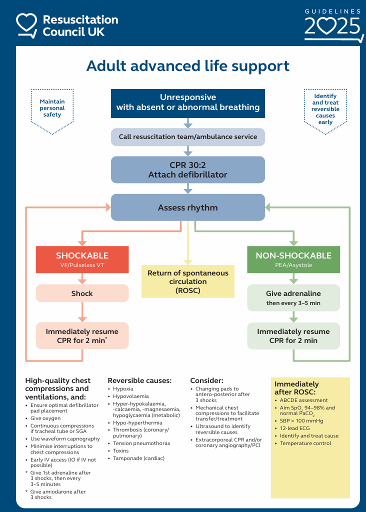
The 2025 Resuscitation Council (UK) Guidelines are included in this CGO for quick reference:


2025 Advanced Life Support Algorithm RCUK


Danger

A dynamic risk-assessment should be conducted on approaching a casualty. Ensure you do not put yourself in danger during resuscitation. Extract the casualty into a space that enables effective intervention, if required. 

Response

Check the patient’s response by a ‘shake and shout’ approach. If they respond, the patient is not in cardiac arrest and should be assessed as appropriate using a systematic approach (MARCH).

Loud Vocal Alarm

Shout for help using a loud vocal alarm (providing this doesn’t put you into danger). The alarm to be used will be context dependent (e.g. ‘Casualty, Casualty, Casualty’ on a maritime platform, ‘Man-Down’ in a land environment etc.)

Airway

Check the patient’s airway. Remove anything visible that is or could obstruct their airway before undertaking a head-tilt/chin-lift airway manoeuvre.

Assess for Signs of Life

While maintaining the airway manoeuvre, look for movement of the chest, listen for breath sounds and feel for breath sounds whilst simultaneously feeling for a pulse. Cardiac arrest is confirmed if the patient has no signs of life or no palpable carotid pulse.


Agonal breathing (slow, irregular and gasping or laboured respirations) is a sign of cardiac arrest, not a sign of life.


Declare Cardiac Arrest

If there are no signs of life declare ‘Cardiac Arrest’ to any team members present. 


Call for Help

Use whatever means are available to call for further assistance. This will be context specific (for example, calling an emergency number in a treatment facility, calling HQ1/control room emergency numbers onboard a ship/submarine, utilising radio communication etc., or ringing the civilian emergency services if in the Firm Base or a civilian setting).

If you are alone and unable to call anyone to assist then it may be necessary to leave the patient in order to get help.


Commence CPR

Commence high-quality CPR immediately:

  • Location: Centre of the chest on the lower half of the sternum.
  • Compression rate: 100–120/min
  • Depth: 5–6 cm
  • Ensure full chest recoil
  • Use basic airway manoeuvres and 15L Oxygen (using self-inflating bag) once available.
  • Ventilation: Delivered over 1 second for each ventilation with enough volume to see chest rise (typically 300-600ml). Avoid excessive ventilation which can worsen outcomes.
  • Ratio of 30 chest compressions to 2 ventilations whenever possible and if safe to do so, otherwise continue compression only CPR.
  • Minimise pauses in chest compressions to deliver the two ventilations. 


Apply Defibrillator

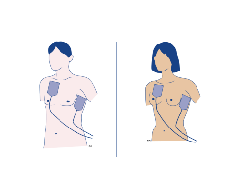
Apply defibrillator pads as soon as available. Pads should be placed initially in an antero-lateral position. Ensure firm contact with clean skin, avoiding excessive hair, sweat or poor adhesion.

2025 Resuscitation Council ALS guidelines highlight that pads are commonly placed in a poor position. Take care to ensure that pad placement is correct. The right (anterior) pad goes below the right collarbone/sternum, and the left (lateral/apical) pad goes below the left armpit on the mid-axillary line.


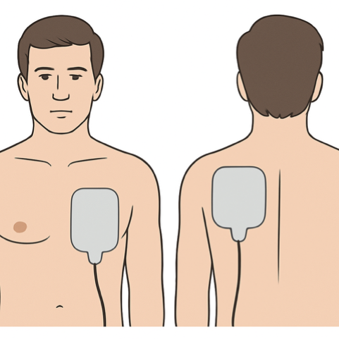
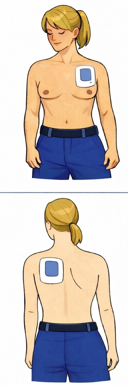
Consider an antero-posterior pad position for vector change defibrillation following three failed shocks in cases of refractory shockable rhythms. The anterior pad is placed to the left of the sternum, avoiding as much breast tissue as possible. The posterior pad is placed at the same height, centred just medial to the left scapula.
Anterior-Lateral Pad Position


Anterior-Posterior Pad Placement Option 1


AP Pad Position Option 2


Use defibrillator in Automated External Defibrillator (AED) mode for initial rhythm analyse and follow audible prompts. If sufficient trained personnel are present, a manual defibrillator should be used.


Ensure oxygen is removed at least 1 metre from the patient unless it is being delivered through an i-gel or endotracheal tube connected to a closed circuit. 


IV/IO access

Obtain Intravenous (IV) or Intraosseous (IO) access. 


Advanced Airway

 Insert supraglotic airway (e.g. iGel) or endotracheal tube (only if trained and competent) with End-Tidal CO2  (wave-form capnography), if available. Once airway is confirmed in place, CPR can be asynchronous (i.e. chest compressions at 100-120 per minute and ventilations at 10 ventilations per minute). If it is not possible to maintain an advanced airway use basic techniques and revert to 30:2 CPR.


Drugs

In a non-shockable rhythm (i.e. if the AED detects a non-shockable rhythm, prompting continuation of CPR without delivering a shock), after recommencing CPR, administer 1mg 1:10,000 Adrenaline via the IV/IO with 20ml 0.9% NaCl or Water for Injection ‘flush’. 

In a shockable rhythm,  give 1mg 1:10,000 Adrenaline via the IV/IO with 20ml 0.9% NaCl or Water for Injection ‘flush’, 300mg Amiodarone IV/IO with a 20ml 0.9% NaCl or Water for Injection ‘flush’ after a third defibrillation shock, with an additional 150mg Amiodarone IV/IO with a 20ml 0.9% NaCl or Water for Injection ‘flush’ after the fifth defibrillation shock. 

Once a dose of adrenaline has been given during cardiac arrest, deliver 1mg of adrenaline via the IV/IO with a 20ml 0.9% NaCl or Water for Injection ‘flush’ every 3-5 minutes (i.e. after every other cycle). 


Minimise interruptions

Do not pause CPR to check for breathing or a pulse unless the AED prompts a reassessment. After a shock is delivered, resume CPR immediately as directed by the AED. Clear signs of life (normal breathing, purposeful movement, palpable pulse detected by a healthcare professional, significant rise in end-tidal CO2) should prompt reassessment. All interruptions to CPR should be minimised and should not take longer than 5 seconds. 


Identify and Treat Reversible Causes

The most frequent reversible causes are referred to by the mnemonic “4Hs and 4Ts”; hypoxia, hypovolaemia, hypo/hyperkalaemia, hypo/hyperthermia, cardiac tamponade, tension pneumothorax, thrombosis and toxins.

See the accordion content section of this CGO for further information and specific treatment recommendations for reversible causes of cardiac arrest

Hypoxia is a very common cause of cardiac arrest with causes including trauma (see Traumatic Cardiac Arrest CGO), airway obstruction, sedation, drowning, blast or smoke inhalation.

Hypovolaemia may be due to absolute volume loss (e.g. haemorrhage) or distributive (e.g. vasodilation and capillary leak during from sepsis or anaphylaxis, hydrostatic pressure effects of prolonged water immersion or neurogenic). 

Hyperkalaemia / Hypokalaemia and Metabolic causes should be considered in all patients. Whilst renal failure is unlikely in Service Personnel, dehydration / rhabdomyolysis due to environmental or physical workload, crush injuries or extensive burns do occur.

Hypothermia or Hyperthermia is more likely than in civilian practice due to the risks of immersion, exposure to high and low temperatures and workload of military personnel. 

Thrombosis encompasses a blood clot or multiple clots causing a coronary vessel occlusion (Myocardial Infarction) or pulmonary embolism. Consider this as a reversible cause, particularly with a sudden unexplained collapse, recent dehydrating activity or environment, patient history suggestive of risk factors or previous clot or if there is evidence of Deep Vein Thrombosis (DVT).

Tension Pneumothorax without trauma is rare, but does occur, particularly in ventilated patients, or those with history of lung disease (e.g. asthma). 

Cardiac Tamponade should rarely be considered outside an inpatient setting post cardiac/mediastinal surgery except in the context of trauma to the neck or torso (particularly penetrating). If suspected, manage as per traumatic cardiac arrest guideline. 

Toxins may be a relevant reversible cause. In a military environment that includes those from a CBRN source. 

See the accordion content section of this CGO for further information and specific treatment recommendations for reversible causes of cardiac arrest


Ceasing resuscitation

Resuscitation can and should be discontinued in specific circumstances. See the accordion content of this CGO and the cessation of resuscitation guideline (link to follow).


Return of Spontaneous Circulation (ROSC)

ROSC marks the next phase in treatment from cardiac arrest. See the accordion content of this CGO for further information. 


In the event of ROSC:

  1. Reassess the patient in a systematic approach (ABCDE or MARCH). 
  2. Identify and treat cause of the cardiac arrest, aiming to prevent further cardiac arrest. 
  3. Aim to restore physiological normality:
    • Maintain a competent airway. Secure an advanced airway when trained personnel are available. 
    • Begin controlled ventilation and oxygenation immediately after ROSC. 
    • Titrate oxygen to oxygen saturations aiming for normal oxygen saturations for the patient (>92% for a patient with no co-morbidities). 
    • Ventilate if not breathing spontaneously, targeted to maintain normal carbon dioxide level (4.5-6.0 kPa), utilising end-tidal CO2 in the absence of the availability of blood gas analysis.
End-tidal CO2 may be used as a surrogate of levels of CO2, recognising it may underestimate PaCO2 in shocked states.


    • Maintain adequate blood pressure to support organ perfusion. Target a systolic blood pressure > 100 mmHg or a mean arterial pressure (MAP) 60-65 mmHg. Use fluids alone if not competent and experienced with use of inotropes or vasopressors. 
    • Ensure continuous ECG monitoring to assess rhythm and detect ischaemia. Patients with ST-elevation on ECG or strong suspicion of cardiac event should be managed using the ACS guidelines. 
    • Check blood glucose level and treat to a normal level. 
    • Check temperature. Actively prevent fever by targeting temperatures to below 37.5⁰C in patients who remain comatose after ROSC. Comatose patients post-ROSC with mild hypothermia (32-36⁰C) should not be actively warmed to achieve normothermia. Temperature should be maintained for 36-72 hours in comatose survivors. 

Advanced Assessment & Management

Management should focus on the interventions that are evidence based to improve outcomes: early defibrillation for shockable rhythms, high-quality chest compressions with minimal interruption, timely drug delivery, early identification and treatment of reversible causes, and structured post-ROSC care.

In deployed environments, this must be delivered alongside clear leadership, parallel tasking, and visible escalation of appropriate care.

Prolonged Casualty Care

Within advanced life support (ALS), Prolonged Casualty Care (PCC) requires a deliberate transition from time-limited algorithmic resuscitation to ongoing physiological stewardship, resource conservation, ethical decision-making, and operational integration. The overarching aim is preservation of life and organ function while maintaining force protection and mission effectiveness. As per the 2025 national guidelines, avoid prognostication before 72 hours after ROSC unless there are clear clinical reasons. 

Across all roles, ALS should be delivered in accordance with contemporary guidance from the RC(UK) adapted proportionately to the operational environment and available resources. PCC does not imply deviation from evidence-based care, but rather contextualised application of that care over extended timelines with constrained resources.

At forward medical locations, PCC is delivered by personnel without the full range of interventions available at higher echelons of care, with limited monitoring, pharmacology, and oxygen reserves. ALS therefore relies on high-quality basic life support, early defibrillation, prompt treatment of reversible causes, and airway management using basic manoeuvres and supraglottic airway devices only. Care should be taken to not escalate care beyond that for which personnel have competence.

Airway management at forward locations during PCC is likely to be limited to supraglottic airway devices (e.g. i-gel) and adjuncts. These devices are appropriate for prolonged use provided their position, seal, and ventilation effectiveness are frequently reassessed. Risks of displacement, aspiration, and gastric insufflation increase over time and require vigilance. Insertion of a fine-bore suction through the port on an iGel can help reduce risk. Because definitive airway control may be unavailable, early evacuation planning following ROSC is essential where prolonged ventilation is anticipated.

Ventilation at forward locations is either accomplished by the patient themselves following ROSC or by manual ventilation. Care must be taken to avoid hyperventilation. Hypocapnia following ROSC is associated with worse neurological outcomes and is a predictable risk in austere settings. Where capnography is available, it should be used continuously; where it is not, ventilation rate and chest rise must be deliberately moderated. The operational objective is normoventilation, recognising that precise PaCO₂ control may not be achievable.

Oxygen management is a defining challenge of PCC at Role 1. Oxygen should initially be titrated to post-ROSC target saturations (typically SpO₂ >92%) where supply permits. Where maintaining these targets would result in premature exhaustion of oxygen reserves, it may be clinically and operationally appropriate to accept lower saturation thresholds in order to preserve oxygen availability over the anticipated duration of care. Deliberate acceptance of permissive hypoxaemia (for example SpO₂ in the high-80s to low-90s) may provide overall patient benefit by sustaining oxygen delivery and avoiding catastrophic deterioration from complete oxygen depletion. Such decisions must be made in consultation with a senior clinician via reach-back where communications allow, explicitly documented, and reviewed regularly.

Circulatory management at forward locations focuses on maintaining coronary and cerebral perfusion with limited means. Following ROSC, hypotension should be treated promptly with fluids and authorised vasoactive strategies within local protocols. Infusion capability may be absent, increasing the risk of haemodynamic instability during prolonged care. This limitation should lower the threshold for evacuation where feasible.

Temperature control is a core PCC task. Hypothermia must be actively prevented at all roles, including in temperate or hot environments. Passive insulation, active warming where available, and minimisation of exposure are essential. Induced hypothermia is not required; the aim is avoidance of hypothermia and prevention of pyrexia.

Metabolic and electrolyte derangements frequently evolve during prolonged care. At Role 1, diagnostic capability may be limited or absent, requiring clinicians to rely on clinical context and arrest aetiology. Hypoglycaemia, hyperkalaemia, and severe acidosis should be anticipated and treated empirically where indicated, recognising that inability to monitor response reinforces the importance of timely evacuation.

Neurological care following ROSC in forward locations is supportive. Sedation options may be limited and neurological assessment confounded by reduced consciousness. Seizures should be treated promptly if observed, but routine prophylaxis is not indicated. Clear documentation of neurological status and trends prior to transfer is essential.

Ethical and operational decision-making is particularly acute during PCC in forward lcoations. Prolonged resuscitation or post-ROSC care may significantly degrade team capability and increase risk to others. Decisions to terminate resuscitation must be clinically defensible, aligned with military medical policy, and clearly communicated. Transparency preserves trust in medical support and supports continued operational effectiveness.

In deployed hospital locations, PCC capability expands on a spectrum to include full critical care. Advanced ventilatory strategies, invasive haemodynamic monitoring, renal support, and formal post-cardiac arrest care pathways are available. Neuroprognostication should be delayed and undertaken in accordance with accepted standards, recognising the confounding effects of sedation, hypothermia, and metabolic disturbance. In civilian practice, this is usually no earlier than 72 hours post ROSC. 

Across all deployed medical capabilities, documentation and handover are critical during PCC. Arrest timelines, airway interventions, drug administration, ROSC time, physiological trends, and key decisions (including resource-driven adaptations) must be clearly recorded. Structured handover between roles reduces error and supports continuity of care across prolonged evacuation chains.

Team and welfare considerations are integral to PCC. Cardiac arrest of a serviceperson has disproportionate psychological impact in deployed settings. Clear communication that care delivered was appropriate to role capability and evidence-based is essential. Debrief and welfare support, such as offering TRIM, are operational necessities, not optional adjuncts.

Paediatric Considerations

Separate guidelines are available to support resuscitation of paediatric patients and also neonatal life support.

Background

Operational Considerations. In the military context, cardiac arrest management must maintain operational credibility. Clear leadership, disciplined communication, visible escalation of appropriate care, and structured debriefing are essential to preserve trust, cohesion, and mission effectiveness. Clinicians should consider the impact of their care on the wider operation.

Advanced Life Support (ALS) is fundamentally an attempt to maintain cerebral and coronary perfusion long enough for defibrillation and correction of reversible causes to succeed. Decades of resuscitation science demonstrate that survival from cardiac arrest is driven primarily by the quality and continuity of chest compressions and timely defibrillation, with drugs and advanced technologies offering incremental benefit at best. This evidence base applies directly to military practice, but its operational implications are magnified when evacuation timelines are prolonged, critical care capability is constrained, and medical events unfold within high-risk environments.

At the centre of modern ALS is not only compression rate and depth, but the proportion of the arrest during which effective chest compressions are delivered. During cardiac arrest, blood flow exists only while the chest is being compressed. Each pause results in a rapid fall in coronary and cerebral perfusion pressure, and several compressions are required to rebuild that pressure. For this reason, the proportion of total arrest time spent actively delivering compressions is strongly associated with return of spontaneous circulation and survival, particularly in shockable rhythms. 

In operational environments, whether maritime platforms, forward medical facilities, austere airfields, or during aeromedical evacuation, interruptions are more likely due to confined spaces, personal protective equipment, environmental hazards, platform movement, or limited personnel. The physiological cost of those interruptions is unchanged, but the margin for recovery may be smaller if definitive post-resuscitation care is delayed. Maintaining a high proportion of time delivering effective chest compressions therefore becomes even more critical in military settings.

Early defibrillation for ventricular fibrillation or pulseless ventricular tachycardia remains the most powerful intervention in ALS. Its effectiveness is closely linked to preceding CPR and minimisation of pauses around shock delivery. Modern evidence emphasises that defibrillation success depends less on technological escalation and more on disciplined execution: charging during ongoing compressions, limiting rhythm check pauses, and immediately resuming compressions after shock delivery. Operationally, this reinforces the importance of equipment accessibility, team rehearsal in constrained environments, and leadership that protects compression continuity.

The role of vasopressors, particularly adrenaline, is defined by large contemporary randomised evidence demonstrating increased return of spontaneous circulation and improved survival to 30 days, but no proportional increase in favourable neurological outcome. This distinction is clinically and operationally significant. Adrenaline improves the likelihood of survival, but not necessarily survival without severe neurological impairment. In deployed settings, where prolonged evacuation or limited intensive care capacity may increase the risk of survival with significant neurological disability, understanding what adrenaline does—and does not—achieve is essential. It remains part of ALS algorithms because it increases survival, but its effect size is modest and must be interpreted realistically.

Similarly, anti-arrhythmic drugs for shock-refractory ventricular fibrillation have not been shown to significantly improve survival to discharge or favourable neurological outcome compared with placebo in large trials. More recent evidence suggests that altering defibrillation strategy in refractory ventricular fibrillation, such as changing shock vectors or using Double Sequential External Defibrillation (DSED) may improve survival in selected systems. However, DSED is not recommended within the 2025 Resuscitation Council (UK) guidelines outside of research. Consultation with the council suggests this is due to limited evidence (early observational studies and a single well-conducted study, the 2022 DOSE-VF trial, having a smaller sample size than planned due to it being halted as a result of the COVID-19 pandemic) and the unknown impact on defibrillators being operating outside of their design. Both vector change and DSED should only be utilised in a well rehearsed team. Where feasible, in well-equipped military medical facility with experienced clinicians, these strategies may be considered, but their benefit depends entirely on preserving compression continuity and avoiding excessive pauses.

Airway management evidence further reinforces the primacy of uninterrupted CPR. Large pragmatic trials comparing supraglottic airway strategies with tracheal intubation have shown no meaningful difference in functional neurological outcomes. In operational contexts—where intubation may be complicated by darkness, noise, reduced manpower, or protective equipment—this evidence supports selecting the airway technique that can be delivered rapidly and reliably without degrading the proportion of time compressions are provided. The most technically advanced airway is of little value if it compromises perfusion by increasing interruptions.

Mechanical chest compression devices have similarly not demonstrated survival advantage when used routinely compared with high-quality manual CPR. Their value in military practice lies in specific logistical circumstances—such as prolonged transport, aeromedical evacuation, or situations with limited personnel—rather than intrinsic superiority. Again, their benefit depends on deployment without prolonged interruption.

Post-resuscitation care has evolved substantially. Large, randomised trials have demonstrated no benefit of routine therapeutic hypothermia compared with controlled normothermia and active fever prevention. The contemporary evidence base supports avoidance of hyperthermia and optimisation of oxygenation, ventilation, haemodynamics, and metabolic parameters as the cornerstone of post-ROSC management. In deployed environments, this is operationally relevant: maintaining normothermia and physiological stability is both evidence-based and achievable, whereas complex cooling protocols may not be.

Whilst Extracorporeal Cardiopulmonary Resuscitation (ECMO) is a useful tool within civilian practice, indicated for patients with witnessed, refractory cardiac arrest, typically with a shockable rhythm and a clear reversible cause, it is generally impractical to deliver on operations. However, it may be considered when operating within countries with a developed ECMO system. 

It is recognised that traumatic cardiac arrest follows a distinct pathophysiological and doctrinal framework and is addressed separately within military guidelines. The ALS evidence discussed here therefore primarily informs management of non-traumatic cardiac arrest on operations, including arrhythmic, hypoxic, immersion-related, or other medical causes.

Beyond its physiological and technical dimensions, cardiac arrest on military operations has a profound impact on the wider team and the collective moral contract between personnel and the medical system that supports them. Sailors, soldiers and aviators must have confidence that if one of their own collapses, every appropriate, evidence-based intervention will be delivered with urgency, competence, and commitment. This belief is fundamental to trust in medical support and to the willingness of individuals to continue operating in high-risk environments.

In this context, the credibility of Advanced Life Support is not derived from indiscriminate escalation, but from clearly recognisable, professional, and determined care that aligns with best available evidence. A well-led resuscitation—characterised by prompt initiation of high-quality CPR, early defibrillation where indicated, timely administration of appropriate drugs, and disciplined team coordination—demonstrates unequivocally that the medical team is doing everything that can reasonably influence outcome. Even when resuscitation is unsuccessful, the visible delivery of ALS to a high standard reassures the wider team that their colleague received full and serious medical care.

Importantly, this reassurance is grounded in evidence-based practice rather than futile or theatrical intervention. Personnel are more likely to retain confidence in medical care when interventions are purposeful, timely, and coherent. The ALS evidence base makes clear that survival is driven primarily by maintaining perfusion—reflected in the proportion of arrest time during which effective chest compressions are delivered—and by timely defibrillation where indicated. Delivering these elements well communicates commitment and competence more convincingly than prolonged escalation that degrades care quality.

Leadership and communication are therefore integral components of ALS on operations. Clear command during the resuscitation, visible coordination of tasks, and calm explanation to non-medical leaders about what is being done reinforce trust. When resuscitation efforts conclude, a clear articulation that all appropriate evidence-based measures were delivered helps the wider team process the outcome and maintain confidence in medical support. This supports operational continuity by reducing uncertainty, moral injury, and loss of trust that could otherwise impair team function.

Taken together, the evidence base for ALS delivers a consistent message across civilian and military contexts. Survival depends predominantly on maintaining perfusion and delivering timely defibrillation, while drugs and advanced interventions offer incremental benefit only when they preserve these fundamentals. In operational environments, where constraints are greater and post-resuscitation care may be delayed, disciplined execution of these core principles is even more critical. Doing everything that is known to work—and doing it well—protects both the individual casualty and the collective effectiveness of the force.

Hypoxia

Hypoxia is a very common cause of cardiac arrest with causes including trauma (see Traumatic Cardiac Arrest guidelines), airway obstruction, sedation, drowning, blast or smoke inhalation. Look for history of cyanosis, poor chest rise, difficult ventilation, facial, neck or chest trauma or deteriorating oxygen saturations prior to cardiac arrest.

Apply maximum level of oxygen available (e.g. 15 litres)

Ensure adequate airway management with airway manoeuvres, adjuncts.

Use a two-person technique for bag-valve mask ventilation to ensure a good seal. 

Insert iGel once CPR in progress, with end-tidal CO2 when available. 

Check for bilateral chest rise and exclude tension pneumothorax early, if found, manage as per traumatic cardiac arrest. 

In deployed hospital facilities, progression to a definitive airway is usually appropriate once skilled personnel and equipment are available, particularly where arrest is prolonged, aspiration risk is high, or controlled ventilation is required.

A cuffed endotracheal tube is generally preferred over a supraglottic airway in this setting. Endotracheal intubation provides a sealed airway with improved protection against aspiration, more reliable delivery of ventilation and airway pressures, and more consistent waveform capnography to confirm tracheal placement, monitor CPR quality, and detect ROSC. These advantages become increasingly important during prolonged resuscitation, patient movement, or delayed evacuation.

Supraglottic airways, including the i-gel, remain appropriate as an initial or bridging airway, particularly if immediate intubation would cause a prolonged interruption to chest compressions or the skillset/monitoring for ETT is not present. In line with Resuscitation Council (UK) (RCUK) guidance, airway interventions must not compromise compression quality, and intubation should only proceed when it can be achieved with minimal interruption. 

Airway management should also include exclusion of obstruction or displacement, suctioning of regurgitated material, and assessment for reversible pulmonary pathology. 

Where bronchospasm or refractory hypoxia persists, escalation to salbutamol (3–20 micrograms/min IV with caution to regularly check potassium), ketamine (0.5–2 mg/kg/hour IV) or adrenaline (0.05–0.5 micrograms/kg/min IV replacing bolus doses) infusions may be required.

Hypovolaemia

In non-traumatic cardiac arrest, hypovolaemia most commonly reflects distributive or relative hypovolaemia, such as sepsis or anaphylaxis, rather than haemorrhage. Management should therefore prioritise early circulatory support alongside treatment of the underlying cause.

Commence fluid resuscitation with appropriate fluid (blood products if available for trauma). 

Consider trauma history (assess for massive external bleeding, flat neck veins particularly in a non-shockable rhythm). Consider neurogenic shock if prior to cardiac arrest history of high-cervical/thoracic spinal injury, history of hypotension with inappropriate bradycardia with warm, dry skin. Manage as per traumatic cardiac arrest with additional use of adrenaline if neurogenic shock suspected.. 

Consider history of allergen exposure, especially with bronchospasm / difficult ventilation / presence of urticarial rash, remove any potential allergen (medication, stings etc.) and ensure adrenaline as per standard cardiac arrest treatment. Care should not focus on antihistamine and steroids are not first line treatment of anaphylaxis.

Consider history or signs of infection such as fever or hypothermia, particularly in a non-shockable rhythm. Give broad spectrum antibiotics if easily available. 

Septic cardiac arrest requires early vasopressor support, prompt antimicrobial therapy, and source control where feasible. 

Anaphylaxis requires immediate adrenaline administration, supported by oxygen, intravenous fluids, and adjunctive therapies in line with national guidance.

Where haemorrhage is suspected, management should follow the traumatic cardiac arrest pathway rather than this guideline.

Hyperkalaemia / Hypokalaemia / Metabolic Causes

Whilst renal failure is unlikely in Service Personnel, dehydration / rhabdomyolysis due to environmental or physical workload, crush injuries or extensive burns do occur. Living in close proximity to others means GI infections can spread and cause electrolyte loss through diarrhoea and vomiting. Massive transfusion can also raise the patient’s potassium load. 

Check BM and treat any low glucose levels. 

Check pre-arrest ECGs/peri-arrest rhythms on rhythm checks for wide QRS, peaked t-waves, sine wave pattern, or history of progressive bradycardia (hyperkalaemia), flattened t-waves, prominent u-waves, ST depression, prolonged QTc, ventricular irritability (ectopics), atrial tachyarrythmias, polymorphic VT or VF (hypokalaemia). 

If hyperkalaemia suspected, give 10ml 10% calcium chloride to stabilise the myocardium. 

Hyperkalaemia should be treated with immediate intravenous calcium (Calcium gluconate 10% 30 mL peripheral IV or Calcium chloride 10% 10 mL central IV/IO) for membrane stabilisation, followed by intracellular potassium shift using insulin with dextrose (10 units Actrapid with either 50ml of 50% or 125ml of 20% dextrose), sodium bicarbonate, and nebulised beta-agonists. Whilst sodium bicarbonate can also promote a small intracellular shift of potassium by correcting metabolic acidaemia, it should be highlighted that this effect is unreliable and clinically modest so should not be used as first line management. 

Hypokalaemia should be corrected cautiously with intravenous potassium (maximum 20 mmol over 10 minutes) with magnesium replacement (5-10 mmol over 10 minutes) where indicated. 

Adrenaline, which is being given as part of immediate management, may shift potassium from the blood into the cells which will support reduction in hyperkalaemia, but conversely contribute to hypokalaemia. 

Urgent transfer to a facility capable of measuring potassium, with consideration of use of insulin/dextrose  for hyperkalaemia, or magnesium and potassium replacement for hypokalaemia if available on route, on advice of a senior clinician if there is a high index of suspicion. 

Other metabolic causes must not be overlooked. Hypoglycaemia requires prompt intravenous dextrose, while hypocalcaemia—particularly following large-volume blood transfusion—should be corrected with calcium replacement. 

Hypothermia / Hyperthermia

Hypothermia or Hyperthermia are more likely than in civilian practice due to the risks of immersion, exposure to high and low temperatures and workload of military personnel. 

Remove wet clothes (do not just open). 

If the patient feels warm or cool to the touch, ensure measurement of core body temperature (using a rectal thermometer if available). 

If core body temperature greater than 40⁰C, actively cool using ice backs to axillae and groin, cooled fluids, water spraying and fanning. Whole-body cold water immersion is not practical during cardiac arrest.

Hypothermia should be actively excluded and managed, and patients should not be declared dead until rewarmed unless injuries are clearly incompatible with life. Core temperature should be measured using a rectal probe, and active rewarming initiated using forced-air warming and warmed intravenous fluids (or blood products if otherwise indicated), alongside ongoing high-quality CPR.

Defibrillation should not be withheld. However, if the core temperature is below 30 °C, no more than three defibrillation attempts should be delivered, with further shocks deferred until rewarming above 30 °C.

Resuscitation drug administration must be modified in hypothermia, in line with RCUK 2025 guidance. When the core temperature is below 30 °C, adrenaline and other resuscitation drugs should be withheld, as drug metabolism is markedly reduced and accumulation may occur. When the core temperature is between 30 °C and 35 °C, resuscitation drugs may be given but at doubled intervals; for example, adrenaline should be administered every 6–10 minutes rather than every 3–5 minutes. Once the core temperature exceeds 35 °C, standard drug dosing and intervals should be resumed.

Hypothermia should be anticipated in maritime, aviation, cold-weather, or prolonged exposure operations and managed in parallel with ALS.


Tension Pneumothorax

Although more commonly associated with trauma, tension pneumothorax remains a recognised reversible cause of arrest in medical patients, particularly during positive-pressure ventilation or in severe underlying lung disease.

Other signs are high pressures required to ventilate the patient, reduced breath sounds unilaterally (although bilateral pneumothorax is possible), resonant percussion note on the affected side and a deviated trachea (late sign). 

Ensure minimum interruptions to CPR if suspected. 

Perform a needle decompression, thoracostomy or chest drain as team skill set allows on the affected side. 

Where suspected, treatment should not be delayed for imaging. Immediate decompression should be performed, followed by definitive chest drainage once ROSC or physiological stability is achieved.

Tamponade (cardiac tamponade)

Cardiac tamponade should be considered in unexplained pulseless electrical activity or refractory shock. Point-of-care ultrasound may be used during ALS where it does not interrupt chest compressions. Pericardiocentesis may be undertaken as a temporising measure or thoracotomy dependent on local capability and the underlying cause. Follow the Traumatic Cardiac Arrest CGO for suspected tamponade in trauma

Toxins and Drug-Related Cardiac Arrest

Toxicological causes should be actively considered in non-traumatic cardiac arrest. Management is antidote-driven and should proceed in parallel with standard ALS. This includes naloxone for opioid toxicity, sodium bicarbonate for sodium-channel blockade, calcium for calcium-channel blocker toxicity, and lipid emulsion therapy in selected refractory cases. Early airway control and ventilation are frequently required. 

Always reconsider your own safety if a toxic cause is considered. 

Give antidote if immediately available such as Naloxone for suspected opioid overdose.

See toxicology CGOs or utilise other available resources to gather further information, for example TOXBASE, particularly unknown poisoning guidance https://www.toxbase.org/4a24ca/globalassets/upload/toxidromes-july-v-2-2024.pdf


Thrombus

Thrombosis encompasses a blood clot or multiple clots causing a coronary vessel occlusion (Myocardial Infarction) or pulmonary embolism. Consider this as a reversible cause, particularly with a sudden unexplained collapse, recent dehydrating activity or environment, patient history suggestive of risk factors or previous clot or if there is evidence of Deep Vein Thrombosis (DVT).  Typically, coronary thrombus is more likely to cause a shockable rhythm compared to pulmonary embolism for which pulseless electrical activity (PEA), a non-shockable rhythm, is more likely. Thrombolysis is only shown to improve outcome to discharge in PE. 

Focus remains on good quality chest compressions and early defibrillation when indicated for both PE and coronary thrombus.  

If PE is suspected, transfer to a facility capable to delivering thrombolysis if the operational context allows.

Coronary Thrombosis. Acute coronary occlusion remains a leading cause of non-traumatic cardiac arrest. Early defibrillation is central where a shockable rhythm is present, with amiodarone used for refractory ventricular arrhythmias. Following ROSC, prompt ECG assessment is required. Where PCI is unavailable, thrombolysis may be considered if suspicion is high and bleeding risk acceptable. Selected Role 3 facilities may consider extracorporeal CPR in line with local governance.

Pulmonary Thrombosis. Massive pulmonary embolism should be suspected in PEA arrest with relevant risk factors. Thrombolysis during cardiac arrest is appropriate where PE is strongly suspected, recognising that prolonged CPR, often 60–90 minutes, may be required following lysis. Give 50mg bolus Alteplase over 1 to 2 minutes and consider a further 50mg after 15 minutes. Alterplase should be mixed with water for injection of 0.9% sodium chloride, but NOT dextrose as it precipitates. In true cardiac arrest, absolute contraindications are relative, but caution is required if the patient has recent major surgery, intracranial pathology, active bleeding or has a trauma mechanism. 


Return of Spontaneous Circulation

In a military operational environment, care after Return of Spontaneous Circulation (ROSC) should prioritise prevention of secondary injury while accounting for resource constraints, evacuation timelines, and the tactical situation. A structured ABCDE reassessment should be undertaken, led by the most suitable senior clinician available, with early consideration of evacuation to a higher echelon of care. Clear documentation and early reach-back to critical care or cardiology support should occur where available, particularly if PCI or advanced organ support may be required.

Airway, breathing and circulation should be optimised pragmatically, with tracheal intubation if not already performed during the cardiac arrest for comatose patients  or with the use of sedative medication for those requiring controlled ventilation. Oxygenation should target normal oxygen saturations (i.e. SpO₂ 94–98%), but in oxygen-limited environments the lower end of this range may be acceptable to preserve supply. Ventilation should aim for normocapnia, guided by waveform capnography and formal blood gasses where available. Circulatory management should target a Mean Arterial Pressure (MAP) ≥65 mmHg (individualised where appropriate), using cautious fluids and vasopressors such as noradrenaline when required. A 12-lead ECG should be performed early, and suspected STEMI should trigger urgent evacuation planning or specialist discussion.

Neuroprotection should focus on avoidance of fever, maintenance of physiological stability, and prevention of secondary brain injury. Active temperature monitoring should be instituted; in austere settings this may rely on simple environmental control and antipyretics rather than formal targeted temperature devices. Glucose should be maintained in a pragmatic normoglycaemic range (approximately 7–10 mmol/L) using point-of-care testing. Sedation should be titrated to prevent agitation or shivering where temperature control is attempted. Clinicians should remain vigilant for seizures and treat clinically apparent events promptly, recognising that EEG monitoring is unlikely to be available forward.

A continued search for reversible causes should occur even after ROSC, with particular attention in military contexts to tension pneumothorax, tamponade, toxin or CBRN exposure, electrolyte disturbance, and pulmonary embolism. Point-of-care ultrasound, if available, should support haemodynamic assessment if a suitably competent user is available. If the patient had a shockable rhythm, consider giving 900mg amiodarone infusion of 24 hours. 

Post-event debriefing should occur when operationally possible to support team learning and psychological wellbeing, maintaining confidence that proportionate, evidence-based care was delivered.

Discontinuing Resuscitation

Resuscitation can be discontinued when:

  • The patient shows clear signs of life i.e. Return of Spontaneous Circulation (ROSC).
  • You are directed to cease resuscitation by a senior clinician, following discussion of all of the circumstances of the case, with no realistic reversible cause identified.
  • You are unable to gain advice by any means of a senior clinician, the patient has had high quality ALS (i.e. effective CPR, all reversible causes have been actively sought and treated, there has been no ROSC at any point, there are no physiological signs of improvement such as improving end-tidal CO2 and the patient has a minimum of 45 minutes of persistent non-shockable rhythm and the team unanimously agree that continuing is not in the patient’s best interests. See Cessation of Cardiac Arrest Management CGO for further details.
  • It becomes unsafe for you to continue. This includes of an evolving situation (for example the spread of fire onboard a ship, nearby kinetic action, concern for an CBRN hazard or severe air turbulence).
  • You become too exhausted to continue (for example as a lone provider, without other personnel who can provide chest compressions). 
     

RCUK 2025 ALS Algorithm

Last reviewed: 10/08/2026

Next review date: 10/08/2027

Evidence method

Consensus guideline